
Page 3 - Carotid and peripheral vascular interventions textbook
P. 3
CHAPTER 10 • RENAL ARTERY INTERVENTION
between the suprarenal aorta and the iliac artery. However, the stenotic kidney. With prolonged exposure, there is
in some instances, the accessory renal artery can have progression of arteriosclerotic lesions and parenchymal
a caliber similar to the main renal artery (Fig. 10-1C), injury to the contralateral kidney that may lead to persistent
thus providing a large part of the renal blood supply. proteinuria and result in pathologic changes from secondary
In this circumstance revascularization of the accessory focal segmental glomerulosclerosis of the contralateral
renal artery stenoses should be performed. Normally, kidney. Revascularization in patients with severe unilateral
a main renal artery remains intact for several centimeters renal artery stenosis may prevent atrophy of the affected
prior to dividing into a variable number of segmental kidney and protect the contralateral kidney.
branches. Early subdivision or bifurcation of the main About 20% of patients have a single functioning
renal artery (Fig. 10-1D) is the second most common solitary kidney or bilateral RAS disease. These worse
anatomic variant, and it makes optimal percutaneous case scenarios create a state of sodium and f uid retention
revascularization more challenging. so that volume-dependent hypertension develops which
then aggravates heart failure symptoms in patients
PATHOPHYSIOLOGY who have impaired left ventricular function and causes
progressive worsening of renal function. These subgroups
Signif cant ARAS is generally a luminal stenosis of ARAS are two of the few absolute indications for
>70% that leads to reduced renal perfusion pressure endovascular revascularization.
and stimulates the renin-angiotensin-aldosterone system
(RAAS) (Fig. 10-2) (15). The net effect of this activation ETIOLOGY
results in sodium retention, peripheral vasoconstriction,
aldosterone secretion, vascular remodeling, inf ammation Renal artery stenosis has many etiologies (Table 10-1).
and triggering of additional vasopressor mechanisms The most common is atherosclerosis that is progression
including endothelin and sympatho-adrenergic pathways of aortic atherosclerotic plaque which affects the
(16,17). proximal segments of the renal arteries and the renal
ARAS has two principle pathophysiological ostia (Fig. 10-3A).
consequences: 1) RAAS activation (in unilateral stenosis) Fibromuscular dysplasia (FMD) is the 2 most frequent
nd
and 2) reduced glomerular f ltration and water and salt etiology and is a nonatherosclerotic, noninf ammatory
excretion (i.e., bilateral artery stenosis or Pickering disorder with unknown etiology that typically affects
syndrome (18) or renal artery stenosis of a solitary kidney). women aged between 15-50 years (19). FMD commonly 251
Although the post-stenotic kidney has less perfusion, the involves on the mid to distal portions of the renal arteries
contralateral kidney experiences glomerular hyperf ltration and causes the angiographic appearance of “string of
and hyperperfusion associated with RAAS activation by beads” aneurysmal appearance (Fig. 10-3B). Contrasting
A B C D
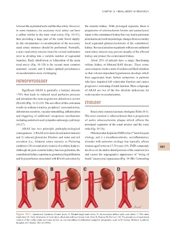
Figure 10-1. Anatomical variations of renal artery. A: Normal single renal artery. B: An accessory inferior pole renal artery. C: Two main
renal artery. D: Early bifurcation of renal artery. (Redrawn with permission from Omar R, Kisansa M, Dehnavi AD. The prevalence of anatomical
variants of the coeliac trunk and renal arteries on contrast-enhanced abdominal computed tomography scans at Dr George Mukhari Academic
Hospital. SA J Radiol. 2021;25:1990.)