
Page 4 - Carotid and peripheral vascular interventions textbook
P. 4
CAROTID AND PERIPHERAL VASCULAR INTERVENTIONS: STEP-BY-STEP
with ARAS, the major clinical manifestation of renovascular transluminal angioplasty (PTA) have resulted in a low
FMD is hypertension which hardly ever causes recurrent threshold for intervening in FMD patients (19).
pulmonary edema or renal impairment. Although medical Takayasu’s arteritis is an uncommon systemic vasculitis
management of hypertension is frequently successful, principally effecting the aorta and its major branches,
the high rates of procedural success, elimination of including the renal artery. Nearly half of the Asian Takayasu’s
hypertension, and low recurrence rate (10%) of percutaneous arteritis patients suffer from renal artery involvement (20).
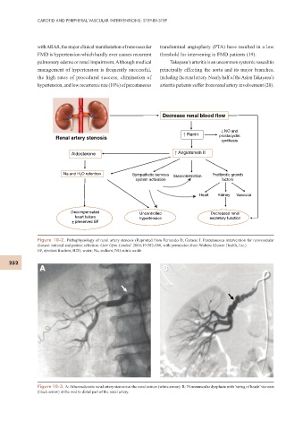
Decrease renal blood flow
! NO and
! Renin prostacyclin
Renal artery stenosis
synthesis
Aldosterone ! Angiotensin II
Na and H 2 O retention Sympathetic nervous Vasoconstriction Profibrotic growth
system activation factors
Heart Kidney Vascular
Decompensated Uncontrolled Decreased renal
heart failure hypertension excretory function
+ preserved EF
Figure 10-2. Pathophysiology of renal artery stenosis (Reprinted from Fernando D, Garasic J. Percutaneous intervention for renovascular
disease: rational and patient selection. Curr Opin Cardiol. 2004;19:582-588, with permission from Wolters Kluwer Health, Inc.).
EF, ejection fraction; H2O, water; Na, sodium; NO, nitric oxide
252
A B
Figure 10-3. A: Atherosclerotic renal artery stenosis at the renal ostium (white arrow). B: Fibromuscular dysplasia with ‘string of beads’ stenosis
(black arrow) at the mid to distal part of the renal artery.