
Page 10 - Carotid and peripheral vascular interventions textbook
P. 10
CAROTID AND PERIPHERAL VASCULAR INTERVENTIONS: STEP-BY-STEP
A B
®
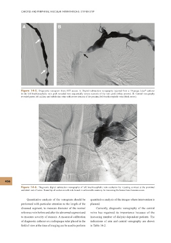
Figure 14-5. Diagnostic venogram from AVF access. A: Digital subtraction venography injected from a 19-gauge Jelco catheter
in the left brachiocephalic vein graft revealed two sequentially severe stenosis of the vein graft (white arrows). B: Central venography
revealed patent left axillary and subclavian veins with severe stenosis of the proximal left brachiocephalic vein (black arrow).
436
Figure 14-6. Diagnostic digital subtraction venography of left brachiocephalic vein occlusion by injecting contrast at the proximal
and distal end of lesion. Round tip of occlusion with side branch is unfavorable anatomy for traversing the lesion from forearm access.
Quantitative analysis of the venogram should be quantitative analysis of the images where intervention is
performed with particular attention to the length of the planned.
diseased segment, to measure diameter of the normal Currently, diagnostic venography of the central
reference vein before and after the abnormal segment and veins has regained its importance because of the
to measure severity of stenosis. A measured calibration increasing number of dialysis-dependent patients. The
of diagnostic catheter or a radiopaque ruler placed in the indications of arm and central venography are shown
f eld of view at the time of imaging can be used to perform in Table 14-2.