
Page 6 - Carotid and peripheral vascular interventions textbook
P. 6
CAROTID AND PERIPHERAL VASCULAR INTERVENTIONS: STEP-BY-STEP
Ipsilateral arm edema usually occurs after a high f ow bleeding during repeated needle cannulation for dialysis,
AV f stula or graft is created in that extremity (Fig. 14-3). can raise the infection risk.
Use of that access for dialysis frequently aggravates SVC syndrome is the severe manifestation of CVD.
the edema more. Swelling, pain and tenderness in the It is distinguished by edema of both upper extremities, the
extremity can resemble cellulitis. Development of tortuous, neck and face with many dilated collaterals across the chest
aneurysmal dilatation of an AVF may exacerbate CVD. and neck. Sometimes, the blood-f ow can be maintained
Prompt AV f stulogram and correction of stenosis can stop via a dilated azygous vein. However, if unrelieved with
progressive deterioration and rupture of the aneurysm. angioplasty or stenting, it may be life threatening and
In chronic CVD, visible, palpable and tortuous veins may result in soft tissue edema of the neck with airway
across the extremity, neck and chest are developed to compression.
divert blood-f ow centrally. Sometimes, the collaterals are
suff ciently large enough to divert blood-f ow to alleviate DIAGNOSIS
the symptoms and signs of CVD, although in most cases
intervention is necessitated. The diagnosis of CVD can frequently be made
Signif cant decline in access blood-f ow, episodes of or suspicious from a thorough history and physical
prolonged bleeding from needle sites following dialysis, examination. Prior central venous catheter implantation
and raised venous pressures during hemodialysis are the history, particularly if of multiple and long duration should
early signs of CVD. Consequently, CVD may reduce warn about the potential for CVD. Presence of pacemaker
access blood-f ow and cause insuff cient dialysis. An AVF or def brillator wires should warrant thorough assessment
generally stays patent even with low blood-f ow, but an for the presence of CVD and its resolution before placing
AVG is more likely to thrombose. Thrombectomy of these an AV f stula or graft on the ipsilateral side. Examination
accesses without attempting to diagnose and treat occult revealing swelling of arm on the ipsilateral side and
CVD can be complicated by worsening of symptoms and many dilated collaterals in the chest or neck indicates
recurrent thrombosis. obstruction to outf ow.
While infection may be a causative factor for CVD, In patients who have not received central venous
CVD can also predispose to infection. In condition of catheterization, other etiologies, such as pacemaker
venous congestion, access thrombosis with excessive wires, thoracic outlet syndrome, hypercoagulopathy, or
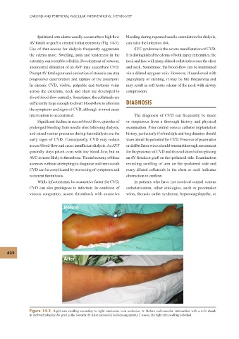
Before
432
After
Figure 14-3. Right arm swelling secondary to right subclavian vein occlusion. A: Before endovascular intervention with a 6-Fr sheath
in the brachiobasilic AV graft at the forearm. B: After successful balloon angiopalsty 2 weeks, the right arm swelling subsided.