
Page 5 - Carotid and peripheral vascular interventions textbook
P. 5
CHAPTER 14 • ENDOVASCULAR TREATMENT OF CENTRAL VENOUS DISEASE
traverses the junction of the clavicle and f rst rib and RISK FACTORS FOR CVS ASSOCIATED
within the anterior-most part of the thoracic outlet. WITH CENTRAL VENOUS CATHETER
Additional to extrinsic compression, repeated forces
in that region often result in f xed intrinsic trauma and CVD is correlated with indwelling intravascular devices
extrinsic scar tissue development. Once primary thrombosis including long-term hemodialysis catheter, pacemaker or
is identif ed, catheter-directed thrombolytic treatment def brillator wires, as well as peripherally inserted central
is normally successful if initiated within 10-14 days of catheter (PICC lines). Risk factors for CVD correlated
clot development, but frequently unmasks an underlying with central venous catheter include:
lesion. Decompression of the venous thoracic outlet, using • Multiple central venous catheter implantations and
thorough external venolysis, resection of the costoclavicular longer catheter dwell times (4,19)
ligament, partial anterior scalenectomy, and f rst rib excision • Subclavian location (4,19,27)
is essential. When bare metal stent (BMS) implantation is • Left sided catheterization (29,30)
conducted on central venous lesions, care should be given • Catheter infection (29,31)
to thoracic outlet syndrome, since they may contribute to • Larger caliber of central venous catheter (12-14 Fr) (32)
complications such as stent distortion and occlusion (24). • Catheter tip position in the proximal part of SVC (33)
Since the occurrence of this syndrome is very seldom and • Catheter composition induced inf ammation
is more frequent in young men in their 20 , so thoracic (e.g., polyethylene and Tef on>polyurethane>
S
outlet syndrome is unlikely to contribute the pathogenesis silicone) (17,34)
of CVD in dialysis patients.
CLINICAL MANIFESTATION
PREVALENCE
Central venous catheter implantation is the most
The incidence of CVD is undef ned and is probably important CVD risk factor. CVD can be totally asymptomatic
underestimated because CVD can be asymptomatic. Serial and can only be discovered by a venogram taken to prepare
or regular venograms also are not normally conducted for AV access implantation (7,15). Following an ipsilateral
following central venous catheter implantation or removal. AV access is created, CVD will probably become symptomatic
Most dialysis patients usually become symptomatic in abruptly because of increased f ow. The symptoms rely
a short time following an ipsilateral AV access is formed upon the particular location of stenosis. While subclavian
as the blood-f ow through the developing dialysis access vein blockage is correlated with edema and venous
rises. The currently available prevalence is limited to the hypertension of the related upper extremity and chest,
studies of symptomatic dialysis patients requiring imaging brachiocephalic vein stenosis impedes blood-f ow from
studies. According to several studies, the occurrence of the same side of the face as well as the upper extremity.
CVD has been published to range between 25% to 40 % Bilateral brachiocephalic vein blockage or SVC blockage
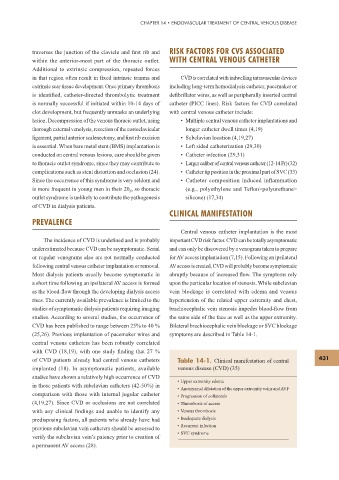
(25,26). Previous implantation of pacemaker wires and symptoms are described in Table 14-1.
central venous catheters has been robustly correlated
with CVD (18,19), with one study f nding that 27 %
of CVD patients already had central venous catheters Table 14-1. Clinical manifestation of central 431
implanted (18). In asymptomatic patients, available venous disease (CVD) (35)
studies have shown a relatively high occurrence of CVD
• Upper extremity edema
in those patients with subclavian catheters (42-50%) in
• Aneurysmal dilatation of the upper extremity veins and AVF
comparison with those with internal jugular catheter • Progression of collaterals
(4,19,27). Since CVD or occlusions are not correlated • Thrombosis of access
with any clinical f ndings and unable to identify any • Venous thrombosis
predisposing factors, all patients who already have had • Inadequate dialysis
previous subclavian vein catheters should be assessed to • Recurrent infection
• SVC syndrome
verify the subclavian vein’s patency prior to creation of
a permanent AV access (28).