
Page 3 - Carotid and peripheral vascular interventions textbook
P. 3
CHAPTER 14 • ENDOVASCULAR TREATMENT OF CENTRAL VENOUS DISEASE
Right internal Left internal
jugular vein jugular vein
Left
Right
subclavian vein subclavian vein
Right
Left
brachiocephalic vein
brachiocephalic vein
SVC
Azygous
vein
Hemiazygous
vein
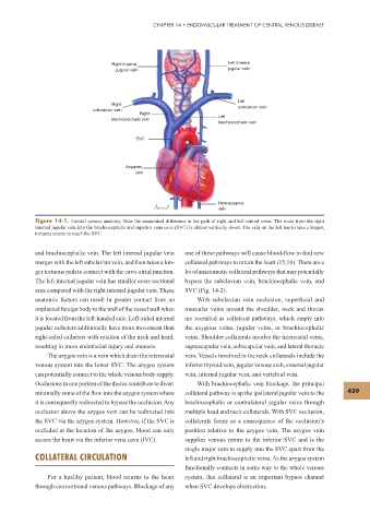
Figure 14-1. Central venous anatomy. Note the anatomical difference in the path of right and left central veins. The route from the right
internal jugular vein into the brachiocephalic and superior vena cava (SVC) is almost vertically down. The vein on the left has to take a longer,
tortuous course to reach the SVC.
and brachiocephalic vein. The left internal jugular vein one of these pathways will cause blood-f ow to f nd new
merges with the left subclavian vein, and then takes a lon- collateral pathways to return the heart (15,16). There are a
ger tortuous path to connect with the cavo-atrial junction. lot of anastomotic collateral pathways that may potentially
The left internal jugular vein has smaller cross-sectional bypass the subclavian vein, brachiocephalic vein, and
area compared with the right internal jugular vein. These SVC (Fig. 14-2).
anatomic factors can result in greater contact from an With subclavian vein occlusion, superf cial and
implanted foreign body to the wall of the vessel wall when muscular veins around the shoulder, neck and thorax
it is located from the left-handed side. Left-sided internal are recruited as collateral pathways, which empty into
jugular catheters additionally have more movement than the azygous veins, jugular veins, or brachiocephalic
right-sided catheters with rotation of the neck and head, veins. Shoulder collaterals involve the intercostal veins,
resulting in more endothelial injury and stenosis. suprascapular vein, subscapular vein, and lateral thoracic
The azygos vein is a vein which drain the intercostal vein. Vessels involved in the neck collaterals include the
venous system into the lower SVC. The azygos system inferior thyroid vein, jugular venous arch, external jugular
can potentially connect to the whole venous body supply. vein, internal jugular vein, and vertebral vein.
Occlusions in one portion of the thorax contribute to divert With brachiocephalic vein blockage, the principal
minimally some of the f ow into the azygos system where collateral pathway is up the ipsilateral jugular vein to the 429
it is consequently redirected to bypass the occlusion. Any brachiocephalic or contralateral jugular veins through
occlusion above the azygos vein can be redirected into multiple head and neck collaterals. With SVC occlusion,
the SVC via the azygos system. However, if the SVC is collaterals forms as a consequence of the occlusion’s
occluded at the location of the azygos, blood can only position relative to the azygos vein. The azygos vein
access the heart via the inferior vena cava (IVC). supplies venous return to the inferior SVC and is the
single major vein to supply into the SVC apart from the
COLLATERAL CIRCULATION left and right brachiocephalic veins. As the azygos system
functionally connects in some way to the whole venous
For a healthy patient, blood returns to the heart system, that collateral is an important bypass channel
through conventional venous pathways. Blockage of any when SVC develops obstruction.