
Page 7 - Carotid and peripheral vascular interventions textbook
P. 7
CHAPTER 14 • ENDOVASCULAR TREATMENT OF CENTRAL VENOUS DISEASE
extrinsic compression of mediastinal veins (e.g., Catheter-based central venography is typically done
mediastinal f brosis, lymphoma, thoracic aortic as an initial procedure to an endovascular intervention.
aneurysm or goitre), should be considered. DSA has more sensitivity than color duplex venous
The initial diagnostic study for CVD is color-f ow ultrasound in the assessment of dialysis access with
duplex venous ultrasound as this technique is noninvasive exceptional ability to discriminate central venous anatomy
with a high sensitivity and specif city (36). A normal vein is from collateral veins. The 2019 Kidney Diease Outcomes
completely compressible on ultrasound. Non compressible Quality Initiative (DOQI) guidelines also recommend
vein with loss of respiratory variations, polyphasic atrial venography before implantation of a permanent AV access
waves, and no doppler f ow augmentation during interrogation in previous subclavian catheterization patients (39).
indicate obstruction downstream of the probe. In addition,
the existence of many neck collaterals generally indicates TREATMENT
CVD. A limitation of this technique is potential acoustic
shadowing from the clavicle, which may impair visualization Treatment options applied for CVD rely upon the
of a short segment of the subclavian vein. It can also be etiology of the disease. Current treatment options include
problematic to visualize central veins using ultrasound in medical therapy, endovascular intervention and open
signif cant muscle mass or obese patients. surgery (i.e., vein bypass). Raising the upper limb and
Computed tomography venography (CTV) and adjunctive anticoagulant treatment may often mitigate
magnetic resonance venography (MRV) are increasingly edema correlated with CVD, particularly when it is involved
utilized alternatives to x-ray contrast venography (37,38), with acute thrombus. However, those measures, are not
which is still the gold standard for diagnosing CVD. applicable in chronic blockage. The use of anticoagulants
MRV is useful in patients with advanced chronic alone has no function in the recanalizing process as the
kidney disease to avoid radiocontrast and to preserve problem remains with the progression of scar tissue. Open
renal function or in those with radiocontrast allergy. CTV is surgical techniques are limited to a few highly morbid
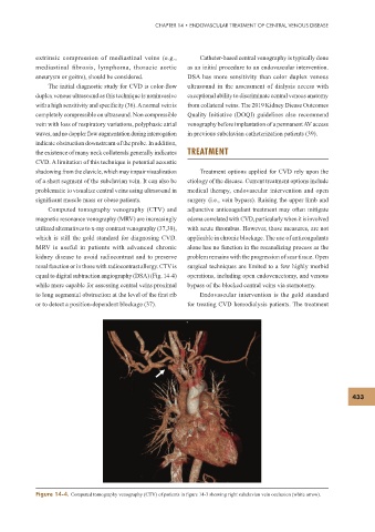
equal to digital subtraction angiography (DSA) (Fig. 14-4) operations, including open endovenectomy, and venous
while more capable for assessing central veins proximal bypass of the blocked central veins via sternotomy.
to long segmental obstruction at the level of the f rst rib Endovascular intervention is the gold standard
or to detect a position-dependent blockage (37). for treating CVD hemodialysis patients. The treatment
433
Figure 14-4. Computed tomography venography (CTV) of patients in f gure 14-3 showing right subclavian vein occlusion (white arrow).